
Dynamique de l'actine et biomécanique de l'embryon précoce
Dynamique de l'actine et biomécanique de l'embryon précoce
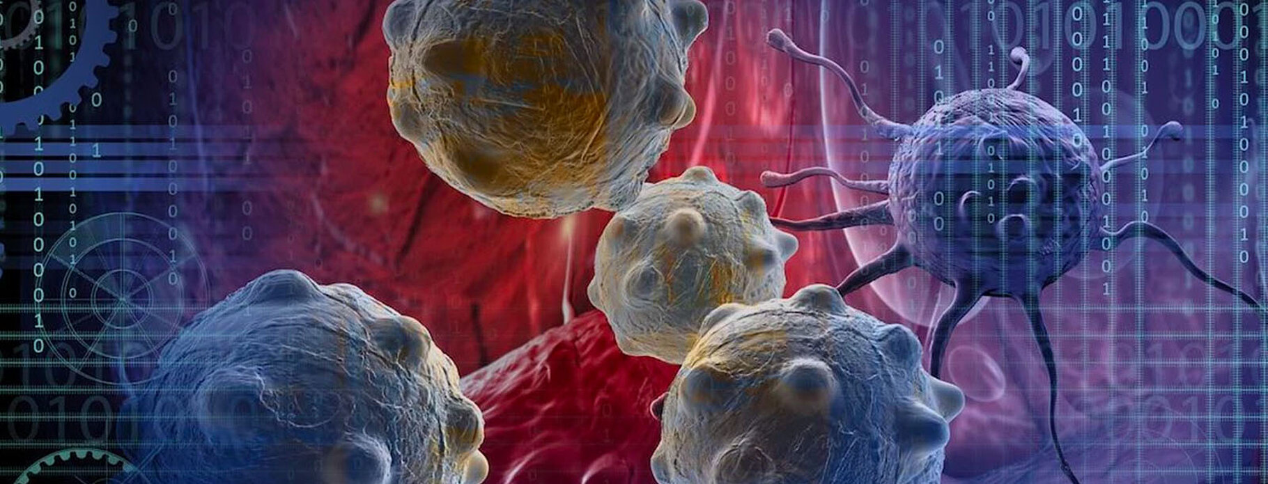
![]() Au cours du développement de l’embryon, la morphologie des cellules est en constant remodelage afin de permettre la bonne tenue des différentes fonctions cellulaires tout en s’intégrant dans son environnent. Les architectures d’actine constituent la structure, ou cytosquelette, donnant forme à la cellule. L’actine définit ainsi en permanence l’architecture interne et externe de la cellule, lui permettant de sentir son environnement et de produire les forces nécessaires pour la motilité ou la divison cellulaire par exemple.
Au cours du développement de l’embryon, la morphologie des cellules est en constant remodelage afin de permettre la bonne tenue des différentes fonctions cellulaires tout en s’intégrant dans son environnent. Les architectures d’actine constituent la structure, ou cytosquelette, donnant forme à la cellule. L’actine définit ainsi en permanence l’architecture interne et externe de la cellule, lui permettant de sentir son environnement et de produire les forces nécessaires pour la motilité ou la divison cellulaire par exemple.
Assemblées, déconstruites ou remodelées en permanence, les architectures d’actines sont diverses et régulées par la dynamique des nombreux acteurs moléculaires qui les composent, tels de véritables blocs de constructions polyvalents. Les réseaux d’actine contrôlent la morphologie de la cellule, sa mécanique, ou même directement l’expression de certains gènes, influencent également l’état de la cellule et ses capacités d’interaction avec l’environnement. Ceci est d’autant plus important dans un organisme en développement, où chacune des différentes cellules doit acquérir une identité et une place spécifique à leur fonction.
Le but de notre recherche est de mieux comprendre comment les dynamiques des architectures d’actine contribuent à l’acquisition de l’identité cellulaire au cours du développement précoce de l’embryon de C. elegans. Pour ce faire nous utilisons des approches interdisciplinaires à l’interface entre biologie cellulaire et biochimie, ainsi que de la microscopie photonique résolutive et des analyses quantitatives. Nous étudions comment les architectures d’actine sont régulées au sein des différentes cellules de l’embryon précoce de C. elegans. Comment certaines particularités peuvent émerger et ainsi impacter les processus d’acquisition des premières identités cellulaires. Notre but étant de comprendre les processus biochimiques de régulation des dynamiques de l’actine in vivo, mais aussi quelles sont les conséquences en cas de perturbation, comme par exemple la présence de mutations génétiques de l’actine reproduisant des maladies rares humaines (actinopathies).

![]()
Membres
Chercheur(euse)s
Doctorant(e)s
Technicien(ne)s
Anciens membres
Laurine Hecht, Etudiante Master1, Université de Strasbroug, 2026
Lysiane Regnard, Etudiante Master1, ESBS / ECPM, Université de Strasbourg, 2026
Eva Schmitt, Etudiante Master1 et Master2, Université de Strasbourg, 2025/2026
Maria Izabella Saad, Etudiante Master2, Université de Beirut, Liban, 2024
Théo Hecquet, PhD, 2024
Fiona Marangoni, Etudiante Master1, Université de Strasbroug, 2024
Alessandro Ulivi, Postdoctoral fellow, 2023
Roxane Benoit, PhD 2024
Anais Goetz, Assistante Ingénieure, 2023
Gregoire Mathonnet, PhD 2023
Caroline Descamps, Etudiante Master2, Université de Mons, Belgique, 2022
Rita Harik, Etudiante Master1, Université de Strasbroug, 2022
Robert Paillaud, Etudiant Master1, Université de Strasbroug, 2022
Delphine Suhner, Assistante Ingénieure, 2021
Pierre Caullet, Etudiant Master1, Université de Strasbroug, 2020
Paul Lettelier, Etudiant BTS, Université de Strasbroug, 2020
Mohamad Alaoud, Etudiant Master1, Université de Strasbroug, 2019
Saurabh Tak, Postdoctoral fellow, 2019
Guillaume Lieb, Etudiant Master1, Université de Strasbroug, 2018
Projets en cours
Project 1 – Actin dynamics and cell identity
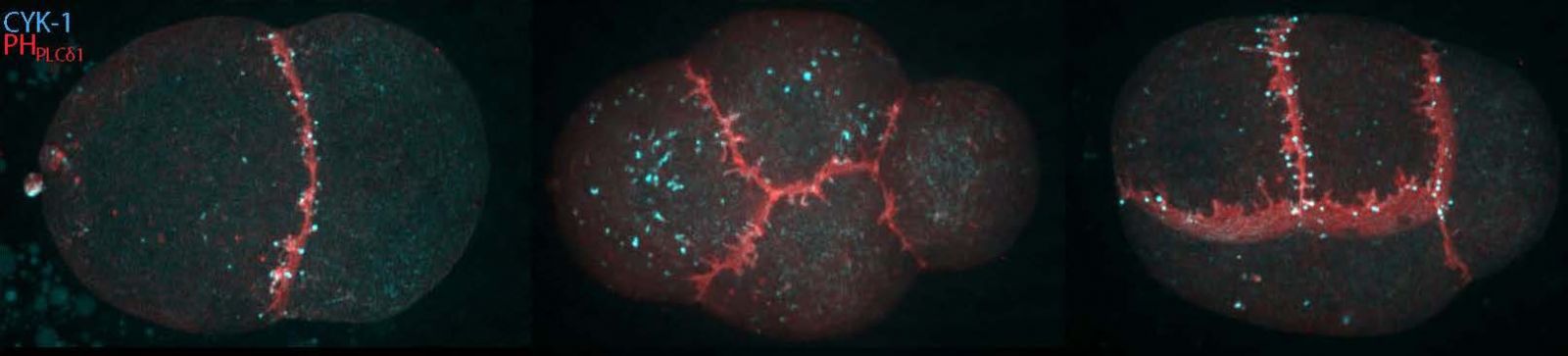
Each embryo experiences enormous challenges in order to successfully transform a passive oocyte loaded with parental material into a polarized embryo of differentiated blastomeres. C. elegans follows a determinate model of development during which invariant cleavage patterns set up reproducible patterns of cell interactions. In addition to rapid volume reduction, asymmetries arise in daughter cells both biochemically and physically, leading in a few divisions to a reproducible but diverse collection of founder cells, the blastomeres.
By the use of in vivo endogenous fluorescent labeling of actin binding proteins (the building blocks) using CRISPR/Cas9 genetic engineering as well as state of the art live imaging techniques and quantitative image analysis, we are studying in the early embryonic cells, the dynamics of individual actin binding proteins and how differences of content or dynamics arise along the lineage.
Our goal is to reveal how actin architectures are spatiotemporally controlled in the different cell types found in the early C. elegans embryo and how some actin specificities could impact cell commitment in the differentiation process during the process of early cell identity acquisition of the blastomeres.
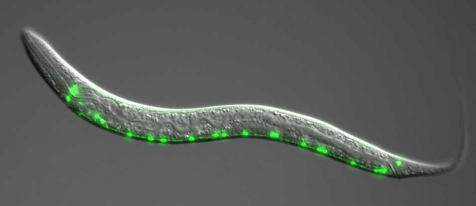
Project 2- CRISPR targeted mutagenesis of C. elegans actin: novel insights into the understanding of human non-muscle actinopathies.
Mutations in the human actin genes ACTB and ACTG1 (beta- and gamma-cytoskeletal actin isoforms) cause a broad spectrum of rare disorders - Non-muscle actinopathies (NMAs)- which show high clinical variability. In human heterozygous loss of ACTB results in a pleotropic neurodevelopmental disorder. Mutations in either nonmuscle actin genes ACTG1 and ACTB were described as a cause of disorders that are now consolidated under the name Baraitser-Winter cerebro-fronto-facial syndrome (BWCFFT).
The five research groups of the European EJP RD PredACTINg consortium have combined their expertise to explore NMA disease mechanisms at a multi-scale level, ranging from single molecules to model organism. The Reymann team is reproducing some actinopathies related human mutations, in the model organism C. elegans using CRISPR/Cas9 mediated genome engineering. These mutants will be used to assess the developmental defects with particular focus on neural development, as well as the perturbation of actin organization and actin dynamics in the early embryonic cells using our fluorescently labelled strains.
The long-term goal of study is to allow a substantial revision of treatment management strategies, provide the basis for future clinical trials and yield broadly applicable functional assays that facilitate genotype-phenotype correlation.
EJP RD PredACTINg consortium: Dr. Med. Di Donato (MHH Hannover, Germany), Prof. Dr. Manstein (MHH, Hannover, Germany), Prof. Dr. Kellermayer (Semmelweis University, Budapest, Hungary), Dr. Bianco (University of Florence, Italy) and Dr. Reymann (IGBMC).
Collaborations et réseaux
Nataliya Di Donato, MHH, Hannovre Allemagne
Johannes Greve, MHH, Hannover, Germany
Francois Robin, Institut de Biologie Paris Seine, Paris, France
Patrick Laurent, Uni Brusselle, Belgique
Laurent Blanchoin, CEA, Grenoble, France
Tri-National DevStemCell Network
GDR Approches Quantitatives du Vivant
Société de Biologie Cellulaire de France
Société Française de Biologie du Développement
Genie, Group of Elegans New Investigators in Europe
Financements et partenaires
- 2023-2025 SeedMoney Proof of Concept funding, IGBMC INRT, ICI platform and AC Reymann
- 2023-2024 Fondation Maladie Rare “Development of experimental models for rare diseases”
- 2024-2027 ANR ADAM, coordinator F. Robin, collaborators A. Jegou, AC Reymann
- 2022-2024 ANR Science avec et pour la société Culture scientifique, technique et industrielle
- 2020-2024 EJP RD, Predacting, European Consortium, coordinatrice Med Dr N. Di Donato.
- 2019-2023 ANR JCJC, DeCaNu.
- 2018-2019 IDEX Grant, Université de Strasbourg.
- 2017-2019 LABEX Start-Up Package, IGBMC, CERBM GIE.

![]()
Publications
2026
Pré-publication, Document de travail
Asymmetric inheritance of actin regulatory machinery shapes cytoskeletal heterogeneity during early Caenorhabditis elegans embryogenesis
- Grégoire Mathonnet
- Roxane Benoit
- Delphine Sunher
- Nadine Arbogast
- Elvire Guiot
- Erwan Grandgirard
- Anne-Cécile Reymann
2025
Article dans une revue
Multiscale characterization of Caenorhabditis elegans mutants to probe functional mechanisms of human actin pathological variants
- Théo Hecquet
- Nadine Arbogast
- Delphine Suhner
- Anaïs Goetz
- Grégory Amann
- Selin Yürekli
- Fiona Marangoni
- Sophie Quintin
- Johannes N Greve
- Nataliya Di Donato
- Anne-Cécile Reymann
iScience ; Volume: 28 ; Page: 113652
Article dans une revue
In vivo detection of ALFA-tagged proteins in C. elegans with a transgenic fluorescent nanobody
- Sophie Quintin
- Maria Izabella Saad
- Grégory Amann
- Anne-Cécile Reymann
microPublication biology
2024
Article dans une revue » Article de synthèse
Acto-myosin clusters as active units shaping living matter
- Karsten Kruse
- Rémi Berthoz
- Luca Barberi
- Anne-Cécile Reymann
- Daniel Riveline
Current Biology ; Volume: 34 ; Page: R1045-R1058
Pré-publication, Document de travail
Classification of human actin pathological variants using C. elegans CRISPR-generated models
- Théo Hecquet
- Nadine Arbogast
- Delphine Suhner
- Anaïs Goetz
- Grégory Amann
- Selin Yürekli
- Fiona Marangoni
- Johannes N Greve
- Nataliya Di Donato
- Anne-Cécile Reymann
2023
Article dans une revue
Meeting report: Third Franco‐Japanese developmental biology meeting “New Frontiers in developmental biology: Celebrating the diversity of life”
- Masayuki Oginuma
- Anne-Cécile Reymann
Genesis - The Journal of Genetics and Development ; Volume: 61
Article dans une revue
The kinesin Kif21b regulates radial migration of cortical projection neurons through a non-canonical function on actin cytoskeleton
- José Rivera Alvarez
- Laure Asselin
- Peggy Tilly
- Roxane Benoit
- Claire Batisse
- Ludovic Richert
- Julien Batisse
- Bastien Morlet
- Florian Levet
- Noémie Schwaller
- Yves Mély
- M. Ruff
- Anne-Cécile Reymann
- Juliette Godin
Cell Reports ; Volume: 42 ; Page: 112744
2022
Article dans une revue
Rapid assembly of a polar network architecture drives efficient actomyosin contractility
- Vlad Costache
- Serena Prigent Garcia
- Camille N Plancke
- Jing Li
- Simon Begnaud
- Shashi Kumar Suman
- Anne-Cécile Reymann
- Taeyoon Kim
- François B Robin
Cell Reports ; Volume: 39 ; Page: 110868
2019
Article dans une revue
Anterior-enriched filopodia create the appearance of asymmetric membrane microdomains in polarizing C. elegans zygotes
- Nisha Hirani
- Rukshala Illukkumbura
- Tom Bland
- Grégoire Mathonnet
- Delphine Suhner
- Anne-Cécile Reymann
- Nathan W Goehring
Journal of Cell Science
Prix/Distinctions
2015, AAAS Newcomb Cleveland Prize, pour ma participation à la publication Chen et al, Science, 2014.
2012, prix de thèse de la Fondation Nanosciences et prix de thèse de l’Université de Grenoble (PRES) pour mon travail en doctorat.