
Mechanics of Oocyte Divisions
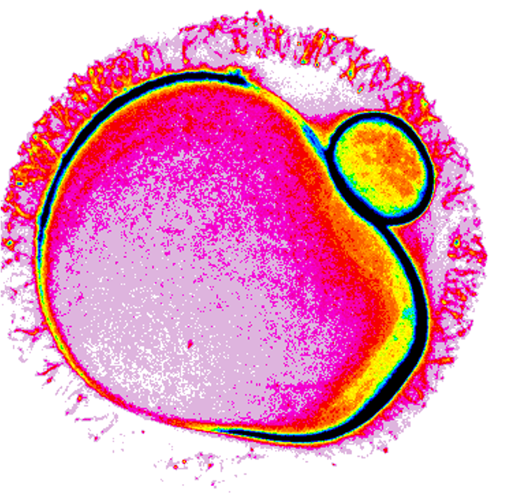
Female meiosis produces oocytes. In mammals, this process ends with two successive asymmetric divisions in size (meiosis I and II), resulting in a large haploid oocyte containing all the maternal stores required for early embryonic development after fertilization. The geometry of these divisions is controlled in part by cortical tension, which decreases in a spatiotemporal manner after meiosis I entry due to actomyosin cortex reorganization, allowing the spindle to migrate along its long axis towards the closest cortex via a Myosin-II dependent pulling mechanism, resulting in an asymmetric division in size. Strikingly, mechanical defects are rather frequent in mammalian oocytes, mirroring the variability in cortical tension and Myosin-II cortical enrichment. In particular, mouse and human oocytes with a too high cortical tension produce embryos that stop developing after fertilization. To elucidate the consequences of excessive cortical tension on oocyte development, we engineered mouse oocytes with excessive cortical contractility by forcing active Myosin-II to their cortex through cortical targeting of the RhoA/ROCK pathway. Our results show that these oocytes progress through meiosis I as successfully as control ones, with comparable timing and geometry of division. However, their cortex spontaneously polarizes upon meiosis I entry, accumulating actin and Myosin-II in a flattened zone opposite as to where division will occur. Directional cytoplasmic flows originate from this polarized zone, pushing the spindle away from it towards the opposite cortex, resulting in the spindle moving faster and in a random orientation. These directional flows disrupt organelle distribution, reducing mitochondrial and Golgi content by half after meiosis I and thus potentially depriving the future embryo of energy reserves and vital organelles. In addition, microvilli and Juno, key players during oocyte fertilization, accumulate precociously and in a restricted manner in the polarized zone, potentially altering fertilization efficiency, which remains to be tested. Finally, we shown that the phenotypes observed in these engineered mouse oocytes exist in natural populations of mouse and human oocytes.
Speaker(s)
Dr Marie-Émilie Terret
Centre interdisciplinaire de recherche en biologie, Collège de France, Paris
France