
Light microscopy
Photonic microscopy

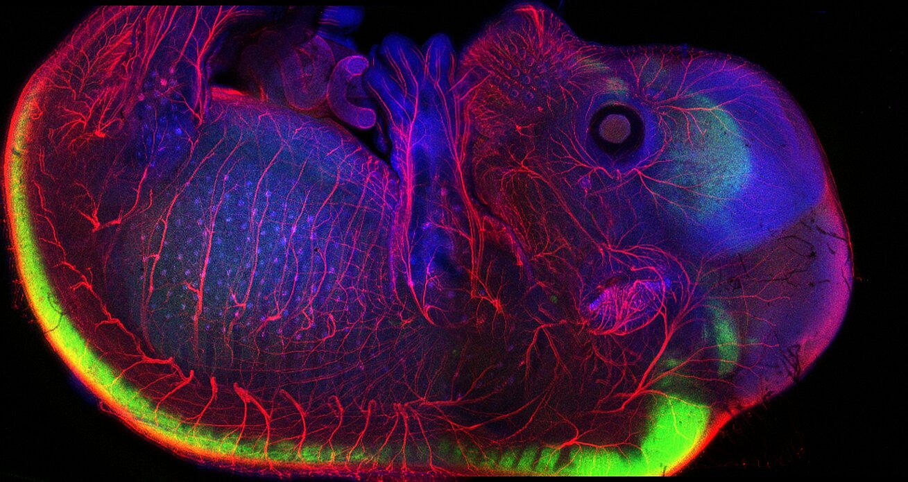
The IGBMC light microscopy core facility, the Imaging Center of the IGBMC (ICI), provides access to cutting-edge optical imaging technologies. Specializing in the study of dynamic biological processes, ICI supports research from the molecular scale to the whole organism. This integrated approach allows for the analysis of study models across multiple size scales, ranging from fine cellular structures to the functioning of organs in vivo.
Next to providing access to microscopes, we offer customized services and training in microscopy as well as bioimage analysis. ICI also collaborates on technological research and development projects together with IGBMC research teams and services.
Do you have a project? Contact us at ici@igbmc.fr. We will get back to you to evaluate the feasibility of your study and to define the optimal imaging or analysis strategy.
Resources
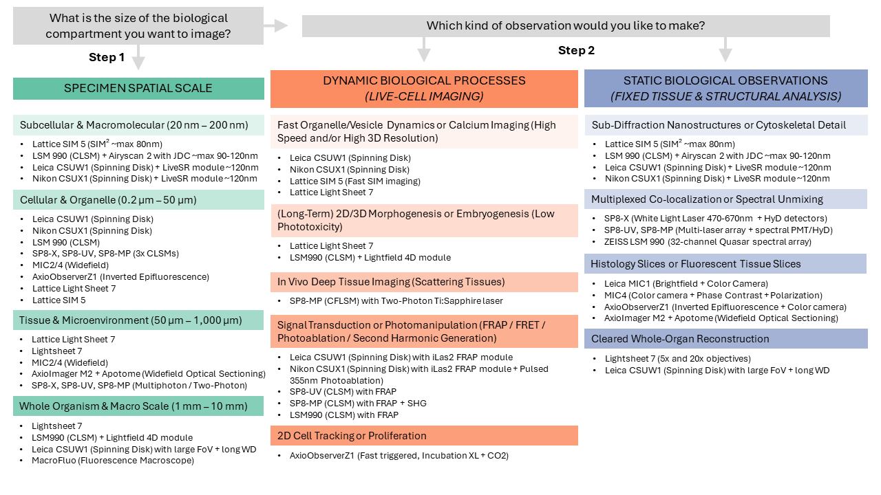
Our facility provides a comprehensive suite of high-end microscopy technologies designed to support diverse research needs, from routine widefield to cutting-edge deep-tissue and super-resolution imaging.
To view detailed technical specifications, access user guides, or browse individual microscope configurations, please visit our OpenIRIS Portal
Our Technology Categories
Confocal & Multiphoton Systems: Designed for high-contrast 3D imaging, deep tissue penetration, and intravital studies. These systems offer precise environmental control and specialized lasers for SHG and photomanipulation.
Super-Resolution & Lattice Systems: These platforms provide sub-organelle resolution and fast volumetric imaging with minimal phototoxicity, ideal for observing live subcellular dynamics.
High-Speed Confocal (Spinning Disk): Optimized for rapid biological processes, these systems feature large fields of view and specialized modules for TIRF, FRAP, and real-time super-resolution of membrane dynamics.
Lightsheet & Widefield Sectioning: Specialized for "gentle" 3D imaging of large fixed samples or cleared tissues, alongside structured illumination systems that remove out-of-focus haze from thick sections (Apotome).
Facility Support & Analysis: Beyond image acquisition, we offer a fully equipped sample preparation suite and high-performance GPU workstations loaded with industry-standard software (including Imaris, QuPath, and Fiji) for advanced data quantification.
Collaborations and networks
The ICI imaging platform is deeply integrated into the scientific community through its active participation in local, national, and international networks. These collaborations ensure our users have access to cutting-edge expertise and state-of-the-art technological developments.
Scientific & Technological Networks
France BioImaging: Member of the national imaging infrastructure via the local node RISEst (Réseau d'Imagerie de Strasbourg et Grand Est).
QuESt Euro-BioImaging GDR Imabio: A national research group facilitating interdisciplinary advances in biological imaging.
Quality Standards & Institutional Certification
IBiSA CoRTecS
