Photonic microscopy
Photonic microscopy
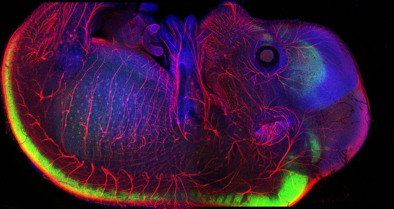
The IGBMC's photonic microscopy platform offers access to state-of-the-art imaging techniques in optical microscopy and specialises in imaging dynamic life processes at the molecular, cellular and whole organism levels. Researchers can analyse, in an integrated approach, their study models at different resolutions, ranging from the finest cellular structures to complex organ function in vivo.
The platform offers services and training in light microscopy and bioimage analysis. The platform collaborates on technological research and development projects with the IGBMC's research teams and platforms and services.
To contact the platform team, send an email to the generic address groupe-mic-photon@igbmc.fr. The platform team will contact you to discuss the feasibility of the project, the best imaging strategy to use and the nature of the services the platform can offer.
Collaborations and networks
The facility is a member of the following network :
- RISE (Réseau d'Imagerie de Strasbourg et région Grand-Est)
- Réseau Technologique de Microscopie photonique de Fluorescence Multidimensionnelle
- GDR-IMABIO
- MIAP
- Network of European BioImage Analysts NEUBIAS
Resources
Confocal and biphotons microscopes
- inverted Leica SP5 equipped with an environmental chamber and a multiphoron laser (680 - 1080 nm) for multiphoton imagign and photoabaltion
- inverted Leica SP8-X equipped white laser and an environmental chamber
- inverted Leica SP8-UV for photoablation, uncaging, DNA damage and photoactivation experiments
- upright Leica SP8-MP equipped with an environmental chamber and a multiphoron laser (690 - 1080 nm) for multiphoton, intravital, photoablation and SHG imaging
Confocal spinning disks
- Yokogawa CSU-W1 (Leica DMi8) equipped with an environmental chamber, FRAP, TIRF 360° and Live-SR super-resolution modules
- Yokogawa CSU-X1 (Nikon TiE) equipped with an environmental chamber, FRAP, photoablation and Live-SR super-resolution modules
Lightsheet microscope
- Leica Digital Lightsheet (restricted to small samples size (<2 mm)) for long term and low phototoxicity live imaging
- Videomicroscopes
- Zeiss Axio Observer Z1 for cell live imaging and medium throughput screen
Widefield Microscopes
- upright microscopes for fluorescence and brightfield
- inverted microscopes for fluorescence and brightfield
Macroscopes
- Leica Macroscope Leica for fluorescence and brightfield
Laser microdissection microscope
- inverted Zeiss Axio Observer Z1 Microscope with PALM MicroBeam microdissection module and environmental chamber
Cell culture and sample preparation
- Cell culture room with cell culture laminar flow hood and cell incubators
- Laboratory space for sample preparation equipepd with a chemical fume hood
Bioimages processinf and analysis
- 4 workstations with a suite of software (Fiji/ImageJ, CellProfiler, Icy, QuPath, Metamorph, Imaris, Matlab)
