
Myotubular myopathy: PI3KC2β, a promising therapeutic target
Myotubular myopathy is a rare genetic disease characterized by severe muscle weakness that can cause breathing difficulties and for which no treatment exists today. In a paper published in the journal JCI Insight, Jocelyn Laporte's laboratory shows that inhibiting the activity of the PI3KC2β enzyme prevents the development of this myopathy in affected mice. Thus, the scientists highlight a promising therapeutic target to treat myotubular myopathy in patients.
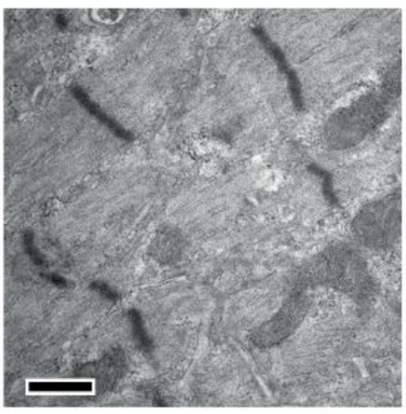
Longitudinal section of mouse muscle model of myotubular myopathy by electron microscopy.
Credit: Messaddeq N, IGBMC
Congenital myopathies are rare genetic diseases characterized by muscle weakness and structural abnormalities of the myofibers, the muscle fibers responsible for muscle contraction. Myotubular myopathy, linked to the X chromosome, is one of the most frequent and severe forms of myopathy in humans. It results from mutations in the MTM1 gene, which codes for a PtdIns3P lipid-targeting phosphatase called myotubularin, a protein required for proper muscle cell function.
Myotubular myopathy is related to a loss of myotubularin and an increase in PtdIns3P levels. Recently, studies show that complete loss of the PtdIns-3-kinase PI3KC2β improves the phenotype of a mouse model of myotubular myopathy.
In this study, the scientists are interested in whether or not PI3KC2β kinase activity is required to improve the phenotype of diseased mice. To do so, they specifically deleted the kinase activity of PI3KC2β while maintaining a normal protein level via a point mutation in the active site, in mouse models of myotubular myopathy associated with loss of the MTM1 gene. The researchers show that suppression of PI3KC2β kinase activity prevents the motor defects, muscle atrophy and weakness, and histological and ultrastructural abnormalities of myotubular myopathy. This prevention correlates with normalization of PtdIns3P levels in muscle, which in turn correlates with normalization of the activity of the mTOR pathway, a pathway involved in many cellular processes.
The scientists highlight that a partial inactivation of the PI3KC2β kinase, allowed only a partial improvement of the phenotype of the mice, highlighting a dose-dependent effect.
These results support the development of specific inhibitors of PI3KC2β kinase activity to improve myotubular myopathy.

Credit: Nattarayan V, IGBMC
Cross sections of mouse muscle stained with succinate dehydrogenase. The labeling stains the mitochondria blue. Labeling is homogeneous in muscle fibers of a control animal (left), whereas mitochondria are abnormally accumulated in the center of smaller muscle fibers (red arrow) in the myotubular myopathy model (center, KO/WT). Inactivation of PI3KC2β kinase activity (right, KO/HO) prevents these abnormalities, and the size of muscle fibers and distribution of mitochondria are similar to those observed in muscle fibers from a control animal. Scale=50µm.