Pathophysiology of Down's syndrome and rare dose-effect diseases causing intellectual disabilities (ID) or autism spectrum disorders (ASD) and other comorbidities
Team Leader : Yann HERAULT
Department : Translational medecine and neurogenetics
SUBGROUP LEADER
 From clinic to basic research
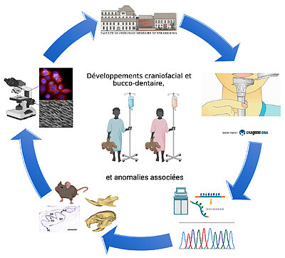
Dental development, which takes place between the 6th week of embryogenesis and 18-25 years of age in humans, is underpinned by epithelial-mesenchymal interactions between the oral ectoderm and the ectomesenchyme from the cephalic neural crests. It crosses all the major developmental signaling pathways (FGF, WNT, SHH, NF-KB, TGF ...) and results in the establishment, according to predefined patterns, of highly specialized mineralized extracellular matrices (enamel, dentin, cementum, alveolar bone). Mineralization fixes these stages in time and allows the presence of developmental anomalies in the form of dental anomalies of number/shape/size/structure/eruption/resorption to be assessed later in children and adults. These abnormalities most often exist in a syndromic context but may appear isolated because of the difficulty of association with other clinical signs. Odontogenesis is part of the dynamics of craniofacial development.
The research work carried out within the team in interaction with the Reference Center for Rare Oral and Dental Diseases of the University Hospitals of Strasbourg and the O-Rares network of the Filière TETECOU and the ERN CRANIO aims at facilitating the understanding of the pathophysiology of these rare diseases and thus reducing the diagnostic wandering of affected persons. They combine genetic approaches: identification of new genes and genetic mechanisms (WGS, WES, RNAseq...), development and transfer of diagnostic tools (databases, GenoDENT NGS Panel, virtual expert AI for diagnostic assistance...), precision of the clinical phenotype, with functional validation approaches using cellular and animal models.